Biology Dissection Images
Magnified insect bodies and specimen close-ups reveal the intricate anatomy found in biology classrooms. 435 images capture the precise detail of beetles, flies, and other organisms under magnification, perfect for educational materials exploring internal and external structure.
Showing 435 of 435 images

pixabay

pixabay

pixabay

pixabay

pixabay

pixabay

pixabay

pixabay

pixabay

pixabay

pixabay

pixabay

pixabay

pixabay
pixabay

pixabay

pixabay

pixabay

pixabay

pixabay
About Biology Dissection Photography
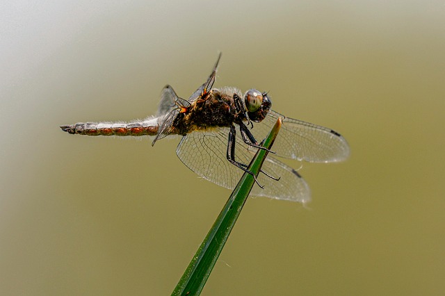
Macro photography dominates the collection, with extreme close-ups of insect exoskeletons, segmented bodies, and fine anatomical features. Mushroom specimens and garden organisms appear alongside microscope imagery, showing both whole-organism and cellular-level perspectives. The visual range spans from individual beetle parts to complete dissected specimens in laboratory settings.
High magnification isolates fine textures—compound eyes, wing veins, and chitinous surfaces—against soft, neutral backgrounds that keep focus on anatomical detail. Natural and controlled lighting creates shadow and depth that emphasizes three-dimensional structure without distraction.
Related Education & Learning Topics
Classroom imagery and student documentation share the same documentary aesthetic, capturing hands-on learning moments with similar lighting and compositional restraint. School laboratory settings use identical macro techniques and close-framing to highlight practical scientific work.
Explore More Free Images
Medical textbook publishers often pair dissection photography with anatomical illustrations to create comparative learning materials. Environmental science documentaries benefit from high-magnification specimen imagery to explain ecological relationships and organism classification.
Download Biology Dissection Images
Educational blog posts explaining insect anatomy rely on these images as header visuals and inline diagrams that break up text-heavy content. Presentation decks for biology courses use macro specimens as full-slide backgrounds, allowing educators to build annotation layers directly over anatomical features.